Retinopathy of Prematurity
What is retinopathy of prematurity?
Retinopathy of prematurity (ROP) is an eye disease where the retina does not mature completely in an infant born prematurely. This can result in severe consequences such as retinal detachment and may result in blindness. However, in recent years there has been great progress in the monitoring and treatment of this eye disease, reducing ROP-related blindness from 50% to 10% in high-risk babies.
This overview will cover the global problem of ROP, who are at risk, how it is diagnosed, the different grades of ROP, as well as the available treatment options.
The global problem of retinopathy of prematurity
It is estimated that there are approximately 15 million babies born prematurely worldwide each year and ROP remains a leading cause of reversible blindness globally. One landmark multicentre study in North America showed that the risk of developing ROP in infants below 1201 grams in weight was 65.8% and 81.6% in those infants weighing less than 1000 grams at birth.
Unfortunately, there are disparities in the outcomes of ROP-associated blindness relative to the socioeconomic environment to which the child is born. Low socioeconomic areas with poor access to intensive neonatal facilities and expertise may account for 40 to 50% of blindness, whereas very low rates of blindness (less than 10%) are reported in high socioeconomic regions.
Who is at risk of retinopathy of prematurity?
The following are several risk factors of ROP:
- Low birth weight
- Younger gestational age
- Supplemental oxygen
- Invasive mechanical ventilation
- Intraventricular haemorrhage
- Total parenteral nutrition
- Thrombocytopaenia
- Hypoglycaemia
Of these, low birth weight and younger gestational age are the most important factors and determine guidance for screening of these infants.
How is retinopathy of prematurity diagnosed?
Rigorous ROP detection programs are crucial for the meticulous screening of diseases. Early detection of diseases allows for early treatment and better visual outcomes, which is why strict protocols are in place in developed countries. A multidisciplinary approach is essential involving cooperative parents, an astute neonatal team, and an experienced senior ophthalmologist to coordinate care.
ROP screening is determined by identifying those neonates who are at risk of developing ROP. Screening programs vary slightly worldwide. In the United Kingdom (UK), current guidelines screen those babies who weigh less than 1251 grams at birth and who were born less than 31 weeks of pregnancy though slightly larger neonates may be eligible.
In North America, the criteria for screening are more than 1500 grams birth weight and born less than 31 weeks of pregnancy. However, infants considered a concern by their neonatologist who fall outside these criteria may also be considered for screening.
Screening differs depending on the centre that is involved in care. Infants that are at risk are highlighted by the neonatal team and prior to examination, their eyes are dilated with specific eye drops. The ophthalmology team usually consists of at least two members, one of which is a senior ophthalmologist who is experienced in examining and treating ROP. A neonatal nurse familiar with the infant is also present.
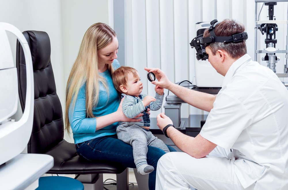
A drop of anaesthetic is instilled into both eyes and a paediatric speculum is used to facilitate examination. The retina is then observed using a special device called an indirect ophthalmoscope. A special tool called an indenter may be used to allow visualisation of the peripheral retina. Some centres also use other forms of imaging with a retinal camera (RetCam or Optos), which is useful for capturing photographs which can then be sent to tertiary centres for an opinion with regards to the treatment. The examination is not painful for the infant but they often express agitation while their eye is being examined. Comfort measures are always encouraged to be used (swaddle, pacifiers, baby toys) and their vital signs are diligently monitored throughout the examination.

Those infants identified as being at risk are closely monitored with serial ophthalmic examinations, usually every 1 to 2 weeks.
Grading of Retinopathy of Prematurity
Systematic international classifications of ROP are in place to allow for standardised diagnosis of the disease. This is based on the location of the disease and its grading.
Grading of ROP is determined by the extent of abnormal vessel growth and occurs in 5 stages (from mild to severe disease):
- Stage I: mild abnormal vessel growth that appears as a white line. Many infants will have complete resolution of the disease without any consequences to their vision.
- Stage II: moderately abnormal vessels that appears as a ridge. Many develop this disease without the need for treatment.
- Stage III: severely abnormal blood vessel growth whereby the blood vessels grow in a different direction (towards the vitreous) instead of along the retina. If associated with tortuous blood vessels “plus disease” this may require treatment to prevent retinal detachment. Treatment is effective if delivered in a timely manner.

- Stage IV: disease that has resulted in a partially detached retina that has been induced by the tractional forces of the scar tissue. This confers a poor prognosis and requires prompt treatment by an experienced vitreoretinal surgeon.
- Stage V: disease that has resulted in a totally detached retina. This can result in complete blindness in an infant which will not improve with age.

Screening programs are implemented to prevent development of stage IV/V of the disease and to intervene as early as required with close monitoring.
Treatment options
Advancements in the treatment of ROP have expanded significantly in the last two decades, resulting in reduced rates of ROP-related blindness in neonatal units with good access to ophthalmic services. Around 90% of babies are in the mild category of ROP and do not require treatment.
Outcomes of treatment emanated from a landmark study which showed that cryotherapy reduced rates of blindness in infants with ROP.
The type of treatment given depends on the stage and location of the disease. The treatment options are:
- Intravitreal medication (an injection that is delivered into the eye to target abnormal blood vessel growth)
- Laser
- Cryotherapy
Surgery (Vitrectomy or scleral buckle)
Intravitreal medication
Intravitreal injections of anti-VEGF (vascular endothelial growth factor) can be delivered in certain circumstances where there is stage III disease closer to the centre of the retina. This can be performed under local anaesthetic (topical drops) in the neonatal unit with the aid of a neonatal nurse, assistant, and experienced ophthalmologist. Risks involved are infection, bleeding, damage to local structures (retinal detachment, cataract), and progression of disease despite treatment. If the latter occurs, they may need further injections or proceed to laser treatment. Often, infants with previous intravitreal injections require longer monitoring compared to those that undergo laser.
Laser and cryotherapy
Laser and cryotherapy work by halting the growth of abnormal blood vessels in the retina that may then lead to retinal detachment. Laser has largely superseded cryotherapy, but the latter remains useful in those where visualisation of the retina may be obscured for example, from a vitreous haemorrhage. Laser treatment usually takes around 45 minutes per eye to perform and often requires an operation theatre set up with an anaesthetist or oral sedation if performed on the ward. The equipment used is very similar to the indirect ophthalmoscope that is used during the screening process. Risks involved include inadvertent central laser burns, cataract, and myopia later in life.
Surgery
Surgery is a last resort option for infants who present with advanced ROP. The goal of screening is always to prevent progression to late stage of the disease. A referral to a tertiary centre with an experienced paediatric vitreoretinal surgeon is required to undergo a procedure called a vitrectomy or scleral buckle. If a vitrectomy is performed, small ports are placed to access the inside of the eye and the vitreous removed. Membranes of scar tissue may be peeled during the surgery and the eye may be filled with a tamponade of gas or oil. If a scleral buckle is performed, the eye is secured with a silicone band that wraps around the eye and allows reattachment of the retina.
It is important that all ROP patients have regular follow-ups with a paediatric ophthalmologist later in life to monitor for amblyopia, strabismus, and refractive error.
DISCLAIMER: THIS WEBSITE DOES NOT PROVIDE MEDICAL ADVICE
The information, including but not limited to, text, graphics, images and other material contained on this website are for informational purposes only. No material on this site is intended to be a substitute for professional medical advice, diagnosis or treatment. Always seek the advice of your physician or other qualified healthcare provider with any questions you may have regarding a medical condition or treatment and before undertaking a new healthcare regimen, and never disregard professional medical advice or delay in seeking it because of something you have read on this website.
References
- “Multicenter trial of cryotherapy for retinopathy of prematurity. One-year outcome–structure and function. Cryotherapy for Retinopathy of Prematurity Cooperative Group,” Arch. Ophthalmol., vol. 108, no. 10, pp. 1408–1416, Oct. 1990.
- J. E. Lawn et al., “Born too soon: care for the preterm baby,” Reprod. Health, vol. 10 Suppl 1, p. S5, Nov. 2013.
- E. A. Palmer et al., “Incidence and Early Course of Retinopathy of Prematurity,” Ophthalmology, vol. 127, no. 4S, pp. S84–S96, Apr. 2020.
- J. AlajbegovicHalimic, D. Zvizdic, E. AlimanovicHalilovic, I. Dodik, and S. Duvnjak, “Risk Factors for Retinopathy of Prematurity in Premature Born Children,” Medical Archives, vol. 69, no. 6. p. 409, 2015. doi: 10.5455/medarh.2015.69.409-413.
- M. F. Chiang et al., “International Classification of Retinopathy of Prematurity, Third Edition,” Ophthalmology, Jul. 2021, doi: 10.1016/j.ophtha.2021.05.031.
- H. A. Mintz-Hittner, K. A. Kennedy, A. Z. Chuang, and BEAT-ROP Cooperative Group, “Efficacy of intravitreal bevacizumab for stage 3+ retinopathy of prematurity,” N. Engl. J. Med., vol. 364, no. 7, pp. 603–615, Feb. 2011.
Tools Designed for Healthier Eyes
Explore our specifically designed products and services backed by eye health professionals to help keep your children safe online and their eyes healthy.
