Pinguecula and Pterygium
Pinguecula and pterygium – Written by Dr Alexis Ceecee Britten-Jones, B Optom (Hons), PhD.
What is a pinguecula and a pterygium?
A pinguecula is a yellow-white growth on the conjunctiva, the clear tissue that covers the white part of the eye. A pinguecula usually grows near the border between the coloured part and white part of the eye, at the 3 and 9 o’clock positions. It is more common for a pinguecula to grow on the side of the eye that is closer to the nose, but it can also grow on the other side, or on both sides.
A pterygium is a larger, fleshy growth that partially covers the cornea, the clear and transparent tissue in front of the eye. A pterygium can start off as a pinguecula, but then grow bigger and cross the border between the white and coloured parts of the eye.

Pinguecula and pterygium are common eye conditions. Neither pinguecula nor pterygium are cancerous, and they do not invade the inside of the eye or spread to any other part of the face or body. Often, the main concern with pinguecula and pterygium is their appearance. However, they can become inflamed or irritating, and affect vision if they grow too large.
This overview will cover how common is pinguecula and pterygium, their causes, who is at risk of developing pinguecula and pterygium, their common signs and symptoms, how they are diagnosed, as well as their treatment strategies.
How many people are affected by pinguecula and pterygium?
Pinguecula and pterygium are more common in certain parts of the world, such as countries located on the equator [1], because people living in these regions have a higher exposure to ultraviolet (UV) light.
Generally, pingueculas are found in 20-40% of people [2, 3], but this proportion has been estimated to be as low as 10% and as high as 75% depending on the country and region being studied [4, 5].
Pterygium is less common than pinguecula [2, 3]. Overall, pterygium is estimated to affect approximately 12% of the world’s population [1, 6]. In Singapore, Malaysia, India, Australia, Indonesia, Korea, and other urban regions, pterygium is found in less than 12% of the population [1, 6-8]. However, in some rural regions in China and Brazil, pterygium has been found in up to 50% of the population [6].
What causes pinguecula and pterygium?
Long-term exposure to the sun or to UV radiation (over five hours a day) is the most common cause of pinguecula and pterygium [9]. This exposure usually takes months and years. UV exposure can be from doing outdoor work; being near environments with reflective surfaces, such as sand, concrete, water or snow; or from tasks such as welding.
Long-term exposure to dry, dusty, and sandy environments is also thought to contribute towards development of pinguecula and pterygium.
Who is more likely to develop pinguecula and pterygium?
People who spend a lot of time outdoors are more likely to develop pinguecula and pterygium.
People with certain occupations, such as factory workers, and machine operators, labourers, and agriculture workers, have a three-fold higher risk of developing pterygium than office workers [10]. Similarly, people who surf regularly are eight times more likely to present with a pterygium than people who do not surf [11].
More men are found to have pinguecula and pterygium than women [1, 7], but this could be because men generally work outdoors for longer periods of time. Smoking and alcohol consumption can also increase the risk of developing pinguecula and pterygium [6].
Pinguecula and pterygium are also more common in older people. In Spain, pinguecula is estimated to be in 20% of people aged between 20-39, 56% of people aged between 40-59, and 73% of people aged over 60 [2].
How does pinguecula and pterygium develop?
UV light breaks down the proteins in the conjunctiva, causing changes in the tissue and excess growth of the fibrous tissue over time.
Pinguecula and pterygium are more commonly found on the side that is closer to the nose because of the way that light focusses when it enters the eye. Light normally enters the eye from the side of the face and concentrates at the tissues closer to the nose. Therefore, the tissues on that side has more UV exposure, and is, therefore, more likely to develop a pinguecula or pterygium.
What are the common signs and symptoms of pinguecula and pterygium?
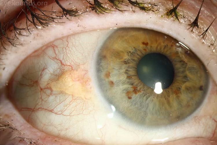
A pinguecula looks like a small, raised, yellow-white growth on the conjunctiva, over the white part of the eye. It can be on one side or both sides of the eye.
A pterygium looks like a triangular, fleshy growth over the front of the eye. It can have thin blood vessels growing over the top. A pterygium appears larger and more obvious than a pinguecula.
Other symptoms and signs that can occur with pinguecula and pterygium are:
- Redness and enlarged blood vessels near the growth
- Irritation and a feeling like there is something in the eye
- Itching, stinging, or burning sensations
- Dry eyes, or a feeling like you have to blink frequently
- Vision changes might occur with pterygium if it’s large
If you experience any of these signs and symptoms, schedule an appointment with an eye health professional to get your eyes checked. It is also important to note that the development of eye conditions may even start before symptoms appear, which makes going for regular and timely eye checks that much more essential.
Different severities of pinguecula and pterygium
Pinguecula normally does not cause any symptoms. Sometimes, they can feel mildly irritating.
Occasionally, a pinguecula can become inflamed, in a condition called pingueculitis, and cause the eyes to become red. Pinguecula will usually not affect vision.
A pterygium, on the other hand, can pull and distort the cornea as it grows. This can affect the way that light enters the eye and causes changes to vision. If a pterygium is advanced and grows too far in, it can also block vision.
Although both pinguecula and pterygium are non-cancerous, it is important to have them checked to ensure that they are not masking other conditions. It is also important to have regular eye examinations to make sure that no other changes are happening in the surrounding tissues [12].
How are pinguecula and pterygium diagnosed?
Pinguecula and pterygium are usually diagnosed by an eye health professional based on the results of a comprehensive eye check. This includes a vision test and an assessment of the health of the eyes, particularly the structures in the front of the eye.
How does an eye health professional confirm that you have pinguecula and pterygium?
The health professional will ask about any symptoms that you might experience and about your eye and medical history. They will also assess your vision to find out if vision is affected.
The eye health professional will examine your eyes using a biomicroscope. This is an instrument that shines a beam of light onto the eyes so that eye structures can be examined under magnification. This technique allows them to examine the appearance of the pinguecula and pterygium closely.
The eye health professional may put an ocular dye in your eyes to look for signs of damaged cells and to show signs of dryness in the front of your eyes. Some common dyes are sodium fluorescein, which is a non-toxic yellow dye, and lissamine green, which is a non-toxic green dye. These dyes will usually disappear within 10 minutes.
How are pinguecula and pterygium treated?
The main treatment for pinguecula and pterygium is to treat dry eye symptoms and reduce irritation. If the pinguecula and pterygium is large or affects vision, they can be surgically removed.
Lubricating eye drops
When there is a raised bump on the eye, the tear film does not spread over the front of the eye evenly. This can cause symptoms of dry eyes, such as dryness, grittiness, or a stinging sensation.
Lubricating eye drops can help tears to spread more evenly over the front of the eye and reduce dry eye symptoms. A liquid eye drop can be used during the day, and a thicker ointment of gel-based eye drop can be used at night.
Anti-inflammatory eye drops
If a pinguecula becomes inflamed, this can be treated with an anti-inflammatory eye drop to reduce the inflammation. Anti-inflammatory eye drops will reduce redness and irritation, but it will not affect or remove the pinguecula.
Surgical removal
Pinguecula and pterygium can be surgically removed to improve cosmesis or to reduce irritation [13]. Because a pinguecula does not grow across the eye as a pterygium can, surgical removal of pinguecula is rare.
Even after surgical removal, 10-30% of pterygium grows back. Larger lesions have a higher chance of growing back [14]. To reduce the chance of reoccurrence, a small piece of tissue from another part of the eye may be used to cover the spot where it was removed. Using this technique, the pterygium does not regrow in as many eyes as when the spot is left bare [15].
Can I prevent pinguecula and pterygium?
Protecting the eyes from prolonged UV exposure is the best way to prevent pterygia and pingueculas from developing and growing. This includes:
- Wearing wide-brimmed hats and close-fitting sunglasses when being outdoors. The best type of sunglasses for preventing pinguecula and pterygium are ones that wrap all the way around the eye to prevent light from entering from the side of the face.
- Avoiding exposure to environmental irritants, such as smoke, dust, wind, and chemical pollutants.
- Wearing appropriate protective eyeglasses in work environments.
DISCLAIMER: THIS WEBSITE DOES NOT PROVIDE MEDICAL ADVICE
The information, include but not limited to, text, graphics, images and other material contained on this website are for informational purposes only. No material on this site is intended to be a substitute for professional medical advice, diagnosis or treatment. Always seek the advice of your physician or other qualified health care provider with any questions you may have regarding a medical condition or treatment and before undertaking a new healthcare regimen, and never disregard professional medical advice or delay in seeking it because of something you have read on this website.
References
- L. Liu, J. Wu, J. Geng, Z. Yuan, and D. Huang, “Geographical prevalence and risk factors for pterygium: a systematic review and meta-analysis,” BMJ open, vol. 3, no. 11, p. e003787, 2013, doi: 10.1136/bmjopen-2013-003787.
- E. Viso, F. Gude, and M. T. Rodríguez-Ares, “Prevalence of pinguecula and pterygium in a general population in Spain,” (in eng), Eye (London, England), vol. 25, no. 3, pp. 350-357, 2011, doi: 10.1038/eye.2010.204.
- A. Fotouhi, H. Hashemi, M. Khabazkhoob, and K. Mohammad, “Prevalence and risk factors of pterygium and pinguecula: the Tehran Eye Study,” Eye, vol. 23, no. 5, pp. 1125-1129, 2009/05/01 2009, doi: 10.1038/eye.2008.200.
- Q. Le, J. Xiang, X. Cui, X. Zhou, and J. Xu, “Prevalence and Associated Factors of Pinguecula in a Rural Population in Shanghai, Eastern China,” Ophthalmic Epidemiology, vol. 22, no. 2, pp. 130-138, 2015/03/04 2015, doi: 10.3109/09286586.2015.1012269.
- J. Panchapakesan, F. Hourihan, and P. Mitchell, “Prevalence of pterygium and pinguecula: The Blue Mountains Eye Study,” Australian and New Zealand Journal of Ophthalmology, vol. 26, no. S1, pp. S2-S5, 1998, doi: https://doi.org/10.1111/j.1442-9071.1998.tb01362.x.
- F. Rezvan, M. Khabazkhoob, E. Hooshmand, A. Yekta, M. Saatchi, and H. Hashemi, “Prevalence and risk factors of pterygium: a systematic review and meta-analysis,” (in eng), Surv Ophthalmol, vol. 63, no. 5, pp. 719-735, Sep-Oct 2018, doi: 10.1016/j.survophthal.2018.03.001.
- H. Cajucom-Uy, L. Tong, T. Y. Wong, W. T. Tay, and S. M. Saw, “The prevalence of and risk factors for pterygium in an urban Malay population: The Singapore Malay Eye Study (SiMES),” Br J Ophthalmol, vol. 94, no. 8, pp. 977-981, 2010, doi: 10.1136/bjo.2008.150847.
- X. L. Fang et al., “Ethnic differences in the incidence of pterygium in a multi-ethnic Asian population: the Singapore Epidemiology of Eye Diseases Study,” Scientific Reports, vol. 11, no. 1, p. 501, 2021/01/12 2021, doi: 10.1038/s41598-020-79920-9.
- C. A. McCarty, C. L. Fu, and H. R. Taylor, “Epidemiology of pterygium in Victoria, Australia,” Br J Ophthalmol, vol. 84, no. 3, pp. 289-292, 2000, doi: 10.1136/bjo.84.3.289.
- T. Y. Wong, P. J. Foster, G. J. Johnson, S. K. Seah, and D. T. Tan, “The prevalence and risk factors for pterygium in an adult Chinese population in Singapore: the Tanjong Pagar survey,” (in eng), Am J Ophthalmol, vol. 131, no. 2, pp. 176-83, Feb 2001, doi: 10.1016/s0002-9394(00)00703-0.
- A. D. Lin, K. u. Miles, and M. V. Brinks, “Prevalence of Pterygia in Hawaii: Examining Cumulative Surfing Hours as a Risk Factor,” Ophthalmic Epidemiology, vol. 23, no. 4, pp. 264-268, 2016/07/03 2016, doi: 10.3109/09286586.2015.1119284.
- J. M. Crewe, T. Threlfall, A. Clark, P. G. Sanfilippo, and D. A. Mackey, “Pterygia are indicators of an increased risk of developing cutaneous melanomas,” Br J Ophthalmol, vol. 102, no. 4, pp. 496-501, 2018, doi: 10.1136/bjophthalmol-2017-310686.
- T. Linaburg, D. Choi, V. Y. Bunya, M. Massaro-Giordano, and C. A. Briceño, “Systematic Review: Effects of Pterygium and Pingueculum on the Ocular Surface and Efficacy of Surgical Excision,” (in eng), Cornea, vol. 40, no. 2, pp. 258-267, Feb 1 2021, doi: 10.1097/ico.0000000000002575.
- S. C. Kaufman, D. S. Jacobs, W. B. Lee, S. X. Deng, M. I. Rosenblatt, and R. M. Shtein, “Options and adjuvants in surgery for pterygium: a report by the American Academy of Ophthalmology,” (in eng), Ophthalmology, vol. 120, no. 1, pp. 201-8, Jan 2013, doi: 10.1016/j.ophtha.2012.06.066.
- E. Clearfield, V. Muthappan, X. Wang, and I. C. Kuo, “Conjunctival autograft for pterygium,” (in eng), Cochrane Database Syst Rev, vol. 2, pp. CD011349-CD011349, 2016, doi: 10.1002/14651858.CD011349.pub2.
Tools Designed for Healthier Eyes
Explore our specifically designed products and services backed by eye health professionals to help keep your children safe online and their eyes healthy.